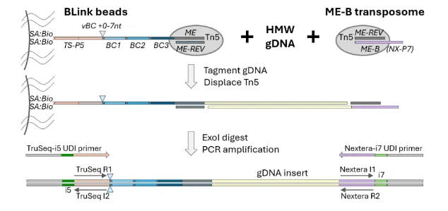
This page provides the BLink-seq library preparation protocol in full as a reference. The official PDF is better formatted for printing and
using in real time at a laboratory bench.
BLink-seq (bead-barcoded linked-read sequencing) technology generates an Illumina library of tagmented inserts that are
individually barcoded based on the molecule of origin. The combinatorial bead-barcode that is added to each sub-
fragment of the original HMW gDNA molecule can be used to infer phasing and linkage of sequenced fragments.
The method uses BLink beads: magnetic streptavidin dynabeads coupled to a 2x-biotinylated oligo containing a TruSeq-
P5 PCR handle, a variable-length barcode (vBC) to offset the start of the Illumina R1 read, a combinatorial barcode (3 x
12nt BC segments), and the Tn5 ME sequence. BLink beads are loaded with purified Tn5 protein to enable on-bead
tagmentation of HMW gDNA. A second Tn5 transposome in solution (ME-B) adds the Nextera-P7 PCR handle. PCR
with Illumina UDI primers (TruSeq-i5 and Nextera-i7) generates the final BLink-seq library.
The final insert size distribution is determined by the frequency of on-bead tagmentation (i5 end) and solution-phase
tagmentation (i7 end). Pilot experiments are recommended to optimize the insert size distribution to maximize the fraction
of the library within the 450-1200bp range. Adjusting both the amount of Tn5 used to load beads (Appendix D) and
especially the amount of solution-phase ME-B transposome is key to optimizing insert size. Once beads are saturated,
adding more Tn5 to beads typically does not alter the insert size, but adding more ME-B transposome during library prep
typically will decrease the insert size distribution. The amount of ME-B transposome required scales with gDNA input.
BLink libraries can be sequenced on Illumina instruments with standard sequencing primers and read lengths.
ME-B transposome: To prepare ME-B transposome in solution, see Appendix B
BLink beads: Can be stored in 3 formats:
fully loaded with active Tn5 transposomes, ready to use: follow protocol below
ME-REV hybridized, ready to load with Tn5: see Appendix D to prepare Tn5-loaded beads
Beads containing barcoded oligos, but requiring final stripping and ME-REV hybridization:
see Appendix C to strip/hybridize ME-REV, and then Appendix D to prepare Tn5-loaded beads
Input genomic DNA: High molecular weight (HMW) genomic DNA will generate BLink-seq libraries with more
linked reads and longer N50 values.
*Once the appropriate amount of ME-B is optimized for a given batch of loaded beads + gDNA samples, ME-B
transposome can be added to the CS+DMF master mix for high throughput library prep, e.g. adding 15μl CS+DMF+ME-
B master mix per sample
Volumes can be modified but the final concentrations of PEG, Triton, CutSmart buffer, and DMF should stay constant.
When increasing the amount of beads per reaction, adjustments to the WASH+PEG buffer concentration can be made. For
example, to use 50μL beads in a final 150ul tagmentation reaction volume, modify the plan to use a 3x formulation of
WASH+PEG buffer and scale up the amount of the CS+DMF master mix to maintain the final concentration per reaction.
The ratio of DNA:beads can change to meet project needs. A higher ratio of DNA to beads will lead to more barcode
clashing because multiple molecules may be tagmented on the same bead and have the same barcode.
Pilot experiments: vary the amount of ME-B transposome to optimize the amount needed for a good final library size
distribution. Typically the primary 2μM ME-B transposome stock is serially diluted in 50% glycerol storage buffer, such
that 1μl of different dilutions are tested in the pilot phase. The optimal amount of ME-B transposome required is related to
the amount of DNA, not the volume of beads; if the volume of beads is increased but DNA stays consistent, do not alter
the amount of ME-B transposome per reaction.
recommended use bench space and equipment that is dedicated to pre-amplification steps through step 5.
1X WASH+T buffer
Prepare fresh each day, store at room temperature
5mL is sufficient for ~20 samples, scale up or prepare more as needed
Volume
Reagent
4.45 mL
Nuclease-free water
500 μL
10x WASH buffer
50 μL
10% Triton X-100
5 mL
Total volume
2x WASH+PEG buffer
Prepare fresh each day, store at room temperature.
Prepare excess buffer, e.g. 1.2 * N samples, this buffer is viscous and may need careful handling and optimization of settings on automated liquid handlers.
Vol/sample
Reagent
13.0 μL
Nuclease-free water
5.0 μL
10x WASH buffer
0.5 μL
10% Triton X-100
6.5 μL
50% PEG8000
25.0 μL
Total volume
HMW sample gDNA
Dilute to target concentration in 1x WASH+T buffer (e.g. 0.3ng/μl in min 5μl).
Rotate (or shake gently < 500RPM) at room temperature to mix well. Do not vortex vigorously to avoid shearing.
Preheat a Thermomixer with a 96well adapter to 55°C
Add 25μl WASH+PEG per sample to labeled strip tubes
Add 5μl Tn5-loaded BLink beads to each tube
Note that beads are not washed before use, especially if ME-B duplex oligo is added when beads are loaded with Tn5 protein (see Appendix C).
Close the strip and flick to mix well
The beads should be evenly dispersed before gDNA is added
One strip at a time, add 5μl diluted gDNA per sample. Immediately close the strip and flick/invert to mix well
Rapid and complete mixing is an important step for assay performance
recommended Use 1x WASH+T buffer as a no-DNA negative control
Incubate minimum 10 min on rotator at room temperature
This pre-incubation step allows DNA to potentially bind to beads before magnesium is added to activate Tn5
Prepare CutSmart/DMF master mix at room temperature:
CutSmart/DMF Master Mix
Vol/sample
Reagent
Notes
4 μL
Nuclease-free water
5 μL
10x rCutSmart buffer
final 1x in tagmentation reaction = 10mM Mg++
5 μL
DMF
final 10% in tagmentation reaction
1 μL
ME-B transposome (e.g. 1:10 dilution of 2μM stock) *
15 μL
Total
*Before the optimal amount of ME-B is determined, do not include ME-B transposome in the master mix.
Prepare excess master mix, e.g. 1.1 * N samples, or more if required for liquid handling dead volume
For large master mixes, a primary stock of 2μM loaded ME-B can also be used; adjust the volume of water accordingly.
Remove the strip-tubes from the rotator. If needed, very briefly pulse-spin the strip tubes to collect the volume at
the bottom of the tube but try not to pellet the beads. Flick gently if the beads are not well dispersed.
one strip at a time add 15μl CutSmart/DMF/ME-B Master Mix per sample. Immediately close the strip and
flick to mix well.
alternative If ME-B is added separately (e.g. pilot phase), first add 1μl diluted ME-B transposome per sample and mix well. Then add 14μl CutSmart/DMF/ME-B Master Mix per sample and immediately mix again.
Incubate samples 30 min, 500RPM on a Thermomixer at 55°C. When finished, proceed immediately with SDS quench.
use a strip-tube reservoir, if needed; excess can be stored at room temperature
When the tagmentation reaction is complete, immediately add 5μl 2% SDS per sample (10% of the tagmentation
reaction volume). Immediately close each strip after adding SDS and flick to mix well
Incubate samples 7 min, 500RPM on a Thermomixer at 55°C
Prepare 10% Triton X-100
use a strip-tube reservoir, if needed; excess can be stored at room temperature
When the SDS incubation is complete, add 5 μl 10% Triton X-100 per sample to quench SDS (equal volume to the amount of 2% SDS added above). Close each strip and flick to mix well.
Pulse-spin each strip and place on a magnetic rack
This is to remove any untagmented oligos on the beads
Prepare ExoI master mix on ice
ExoI Master Mix
Vol/sample
Reagent
17 μL
Nuclease-free water
2 μL
10x rCutSmart buffer (NEB)
1 μL
Thermolabile ExoI (NEB)
20 μL
Total
Prepare excess master mix, e.g. 1.1 * N samples, or more if required for liquid handling dead volume
Prepare a multichannel reservoir with 1xWASH+T buffer
Remove the supernatant from the beads (on magnet) and discard. On magnet, add 100μl WASH+T
optional If desired, beads can be ‘downsampled’ to generate a library for only a portion of the bead-bound
tagmented fragments. Downsampling will generate a library with fewer fragments and lower complexity, but can
increase the percent of linked reads at lower sequencing depths. The remaining tagmented beads can be stored
at 4°C and processed later if desired.
To downsample beads: remove the strip from the magnet, flick or mix to disperse the beads, and transfer the desired portion to a new strip or tube and return to magnet. The equivalent downsampling can also be done at the wash step after ExoI treatment and before PCR.
Remove the supernatant from the beads (on magnet) and discard
If processing many strips, either close them to prevent the beads from drying out while washing all of the strips, or add the ExoI master mix to each strip before continuing with the next strip.
Remove the strip from the magnet and add 20 μl ExoI master mix to each sample. Close the strip and flick to mix. Pulse-spin very briefly to ensure all of the liquid is at the bottom. The beads should remain fully dispersed.
Incubate samples in a thermoscycler: 10min at 37°C, then 5min at 65°C (heat kill)
The last part of the protocol is to attach Universal Dual Index (UDI) adapters/indices to
the DNA via PCR. After this, all that’s left is a final bead cleanup.
Thaw hybrid UDI stocks (mix of TruSeq-i5 + Nextera-i7, 5nM each) on ice. Pulse-spin when thawed.
Prepare PCR master mix on ice with excess, e.g. 1.1 * N samples, or more if required for liquid handling dead volume.
Program a thermocycler as indicated below, including an initial pause step at 72°C. Start the thermocycler so that the block pre-heats to 72°C.
When the ExoI incubation is complete, pulse-spin each strip and place on a magnetic rack.
The samples are stable on the magnetic rack at room temperature.
Set a (multichannel) pipet to 100ul. One strip at a time on magnet, add 100μl WASH+T (no need to remove the
ExoI reaction first – just add WASH+T to the tube). If the beads come off the magnet, pause until the supernatant
is clear. With the same tips, remove the entire supernatant (~120 ul).
Optional: Bead Downsampling
To downsample beads, remove the strip from the magnet after adding WASH+T buffer, and mix to fully disperse
the beads. Transfer the desired volume to a new strip and place on a magnetic rack. When the supernatant is clear,
remove the supernatant. The remaining tagmented beads can be stored at 4° C and processed later if desired.
Move the strips containing the bead pellets to a chiller block or a rack on ice.
On a chiller block or on ice, add 20 μl 2x HiFi Master Mix per sample.
On a chiller block or on ice, add 5 μl hybrid UDI stock (TruSeq-i5 + Nextera-i7, 5nM each) to each sample.
Thermocycler Program
Initial pause: 72°C preheat and hold at temperature
Initial incubation: 72°C for 5min extend tagmentation gaps
Initial denaturation: 98°C for 30sec
12* Cycles of:
98°C for 10sec
65°C for 30sec
72°C for 1min
Final extension: 72°C for 5min
*PCR cycle number may need to be adjusted based on beads and DNA input.
Recommended put away all pre-PCR reagents before moving to a different bench for post-PCR cleanup.
Set up new strips with 22.5 μl DNA cleanup beads (0.9x)
Transfer PCR reaction to new strips containing DNA cleanup beads
Vortex to mix well, incubate at least 5min at room temperature
Pulse-spin and transfer strip to magnet
On magnet, wash 2x with 200 μl fresh 80% ethanol
For each wash, pipette up and down (on magnet) to rinse the beads, then leave the ethanol wash on the beads for minimum 30sec
After 2nd wash: pulse spin and remove residual wash
Allow beads to dry briefly, only until droplets are gone (beads should still be dark and glossy)
Remove from magnetic rack and resuspend beads in 23μl 0.1x TE
Incubate at least 5 min at room temperature, transfer to magnetic rack until clear
Transfer supernatant to a clean, labelled strip tube or plate. This is the final BLink-seq library
Optional: quantify and check the size distribution for all or a subset of samples
Prepare tubes for Qubit assay: add 198 μl 1x HS dsDNA Qubit reagent, hold in the dark at room temperature until
equilibrated.
Transfer 2μl final library stock to Qubit tubes. Vortex, pulse-spin, and incubate 2 min at room temperature.
Read and record Qubit measurements.
Prepare aliquots for size QC, dilute if needed to be in the target concentration range.
Final library size can be estimated by gel electrophoresis or using an instrument such as the Fragment Analyzer (Agilent)
or equivalent. A successful library will typically have a size range from ~300-1500bp, with 40% or more in the target size
range of 450-1200bp. To adjust the size range in the next experiment, alter the amount of ME-B transposome used (more
ME-B transposome = smaller insert sizes and vice versa). Libraries with less material in the target size range can still be
sequenced, but the amount of linked reads may be lower and/or the insert size may be smaller than ideal (<450bp).
Pool final libraries, based on QC metrics (Qubit concentration and size distribution) when available. If the final pool
will be size-selected, plan the pool based on molarity estimates for the target size range.
Recommended: size-select the final pool (450-1200bp) on a Pippin instrument (Sage Science) in order to optimize
insert sizes prior to sequencing.
Sequence on an Illumina sequencer, recommended 2x150 PE reads with dual index reads. Index read lengths should
match the UDI primers used.
To process your BLink-seq data, use Harpy.
Harpy includes several modules for preprocessing reads, aligning reads, calling variants (SNPs and SVs), and more.